Home » Health News »
SARS-CoV-2 interacts with sialic acid cell surface glycans
Severe acute respiratory syndrome coronavirus 2 (SARS-CoV-2) and the earlier SARS-CoV-1 gain entry to host cells by interacting with the ACE2 receptor, while the closely related MERS-CoV enters human cells by binding with dipeptidyl-peptidase.
Besides these major receptors each virus also interacts with a number of additional surface proteins or glycans that aid in initial adhesion and subsequent internalization.
SARS-CoV-2, for example, is known to involve cell surface protease TMPRSS2 to cleave a section of its own spike protein to facilitate cell entry, and a number of other potential receptors have recently been described, including kringle containing transmembrane protein 1 and asialoglycoprotein receptor 1.
Similarly, MERS-CoV is known to interact with some sialic acids, sugar molecules found on the surface of cells, which have also been suggested as a candidate SARS-CoV-2 receptor. Sialic acids are usually found in the terminal position on a variety of glycoconjugates, on the surface of a wide variety of cells, and are also known to mediate the attachment of other viruses such as influenza.
In a paper recently uploaded to the journal Frontiers in Chemistry by Li et al. (July 22nd, 2021) the interaction between the SARS-CoV-2 spike protein and N-acetylneuraminic acid (NANA), the most commonly found sialic acid in humans, is investigated, finding that the molecule may act as a secondary or supplementary receptor to the virus.
How was the study performed?
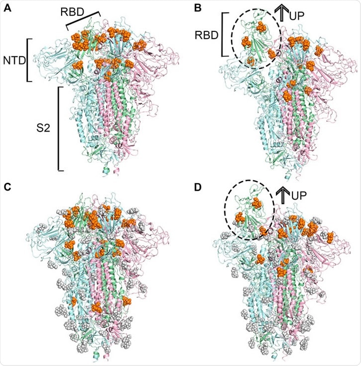
The SARS-CoV-2 spike protein receptor-binding domain (RBD) was modeled computationally, in both open and closed conformations and with and without glycosylation, and each examined for potential binding sites for NANA. When in the closed state, the RBD bore 21 potential binding sites when non-glycosylated and 23 when glycosylated, while the open state RBD had 15 and 17, respectively, totaling 40 unique sites.
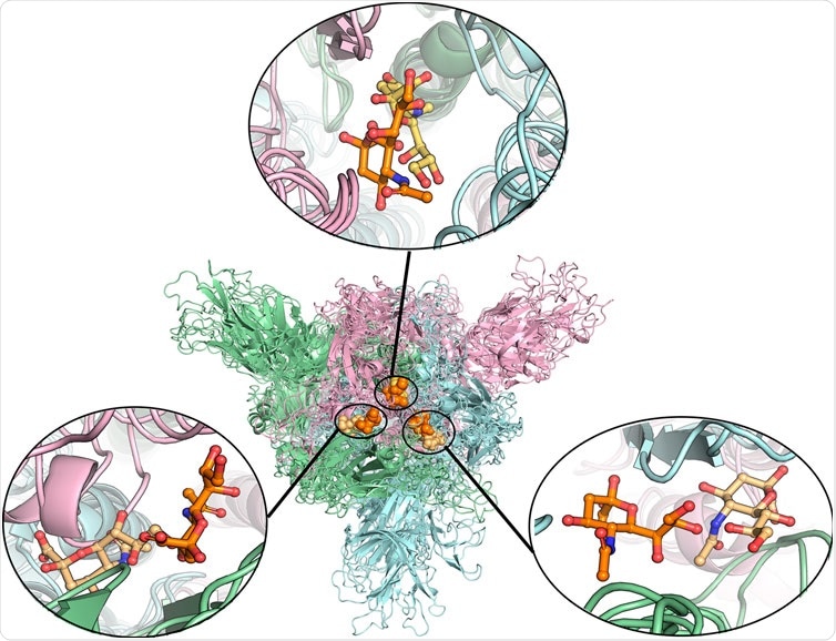
The stability of each binding site was assessed in 20 nanosecond long simulations, finding overall that glycosylation was beneficial to bond stability. Longer 200 ns simulations were performed for the best candidates, finding a number of appealing sites.
The group notes that while in the closed conformation, an additional binding site that spans the RBD is available, being unsuitable when in the open conformation.
The major binding site of sialic acids to MERS-CoV is within the N-terminal domain of the virus's spike protein, while in the case of SARS-CoV-2, the group detected no binding sites here, only within the RBD.
Once bound to a host cell, the spike protein of SARS-CoV-2 undergoes several conformational changes that allow it to pass the cell membrane, some of which are mediated by molecules binding to the protein at particular sites.
The group suggests that the examined sialic acid may potentially be involved in inducing such changes, as one of the major binding pockets identified overlaps with one thought to be involved in conformational changes. However, this has yet to be confirmed.
The identification of this binding site may facilitate drug design efforts, wherein a drug with a similar structure or with a sialic acid targeting group could potentially interfere with the SARS-CoV-2 cell entry process.
- Li Bingqian, Wang Lin, Ge Huan, Zhang Xianglei, Ren Penxuan, Guo Yu, Chen Wuyan, Li Jie, Zhu Wei, Chen Wenzhang, Zhu Lili, Bai Fang, Identification of Potential Binding Sites of Sialic Acids on the RBD Domain of SARS-CoV-2 Spike Protein, Frontiers in Chemistry, DOI=10.3389/fchem.2021.659764, https://www.frontiersin.org/article/10.3389/fchem.2021.659764
Posted in: Medical Science News | Medical Research News | Disease/Infection News
Tags: ACE2, Cell, Cell Membrane, Coronavirus, Coronavirus Disease COVID-19, Glycans, Glycosylation, Influenza, Membrane, MERS-CoV, Molecule, Protein, Receptor, Respiratory, SARS, SARS-CoV-2, Severe Acute Respiratory, Severe Acute Respiratory Syndrome, Spike Protein, Syndrome, Virus

Written by
Michael Greenwood
Michael graduated from Manchester Metropolitan University with a B.Sc. in Chemistry in 2014, where he majored in organic, inorganic, physical and analytical chemistry. He is currently completing a Ph.D. on the design and production of gold nanoparticles able to act as multimodal anticancer agents, being both drug delivery platforms and radiation dose enhancers.
Source: Read Full Article


