Home » Health News »
Artificial Intelligence used to enhance decision making during colorectal cancer surgery for first time

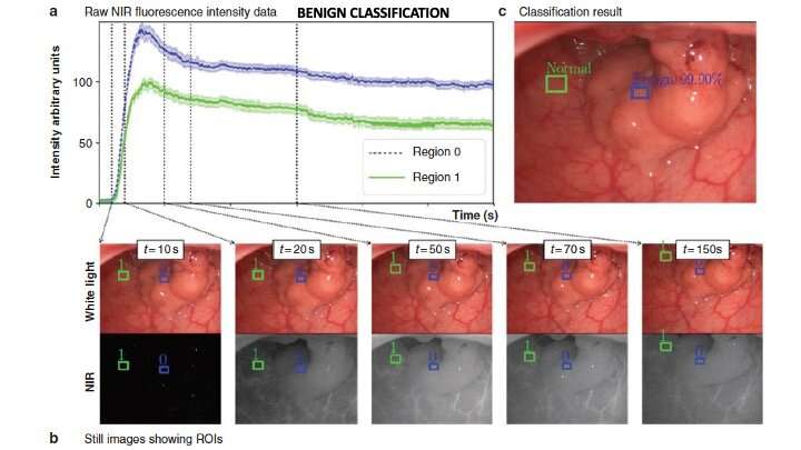
A clinical research study published in the British Journal of Surgery shows that fluorescence guidance (where a fluorescent chemical compound re-emits light with a long wavelength) can enable a colorectal surgeon to assess cancer tissues visually and with more specificity in real-time during surgery, by using near-infrared (NIR) light from an administered fluorophore in conjunction with artificial intelligence (AI) methods.
In this study, supported by the Disruptive Technologies and Innovation Fund 2018, videos from 24 patient (11 with cancer) surgeries were studied. Numerous ROIs (Regions of Interest) from each area of abnormality were selected for analysis from each video. NIR intensities were extracted by tracking the ROIs within each video, focusing on the initial wash-in period. The data set used for analysis included 435 ROI profiles each with 12 perfusion-characterizing features with balanced outcomes. At patient level, the system correctly diagnosed 19 of 20 cancers (95%.)
Prof Ronan Cahill, professor of surgery at University College Dublin (UCD) and the Mater Misericordiae University Hospital (MMUH), said “Surgery has the substantial role to play in the therapy of over two-thirds of all cancers and key surgical decisions are traditionally made by human visual judgements, which assume a static biological FOV (Field of View) during the time frame of the observation (which in surgery is moments).”
“The process for uptake and release of an external substance, such as drugs and contrast agents, are unique in cancerous tissues. As such, we envisaged that an approach combining biophysics-inspired modeling and AI could analyze intraoperative changes in NIR intensities over time in varied tissue, enabling clinically useful lesion classification with high specificity. To translate this knowledge for the first time into an intraoperative surgical decision support tool, a computer vision-AI real time tissue-tracking and categorizing protype has been developed. As the prototype relies only on the NIR fluorescence data stream, it is usable with commercially available imaging systems.”
Source: Read Full Article


