Home » Health News »
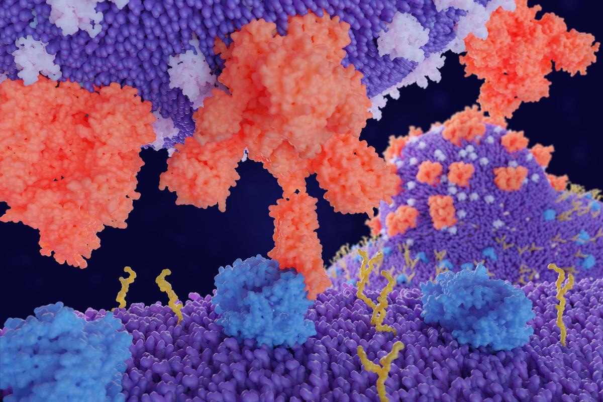
Comparison of RBD/ACE2 receptor complex in wild type SARS-CoV-2 and Omicron variant
An essential component of severe acute respiratory syndrome coronavirus 2 (SARS-CoV-2) is the spike protein’s receptor-binding domain (RBD). The RBD binds with human angiotensin-converting enzyme 2 (ACE2) receptor, giving it entry to infect cells. Most mutations from variants of concern such as Alpha and Beta have reported mutation on receptor binding domain residues. Some antiviral coronavirus treatments work to block the interaction between ACE2 and the RBD, but the Omicron variant and its suspected increased infectiousness may compromise those efforts.
 Study: Scanning the RBD-ACE2 molecular interactions in Omicron variant. Image Credit: Juan Gaertner/Shutterstock
Background
South African scientists identified the Omicron variant in late November. It is estimated to have 32 mutations on the spike protein compared to the 13 to 17 mutations on the Delta variant. Early data shows it is more transmissible than other SARS-CoV-2 variants and can weaken vaccine effectiveness. There is an increase in breakthrough infections from fully vaccinated people, but a third dose or booster shot may provide considerable protection against Omicron.
New Omicron cases are being detected worldwide, with a winter surge observed in South Africa and the United Kingdom. Scientists have predicted that Omicron will overtake Delta as the dominant variant in the United States by mid-January.
New research published in the bioRxiv* preprint server details the molecular landscape of Omicron’s RBD and how it differs from the original SARS-CoV-2 strain. They found the Omicron variant has stronger binding energy with ACE2 and forms a more stable protein complex than prior SARS-CoV-2 strains. The findings could explain why Omicron has a high rate of transmission.
The study
Considering the lack of data available on the Omicron variant’s structure, the researchers used the RBD/ACE2 complex from the original SARS-CoV-2 strain as a template to simulate different models that could explain Omicron’s protein interactions. One thing to note is that the researchers intentionally excluded glycans in the model because previous research suggested that the RBD contained fewer glycans than the rest of the spike protein.
The RBD is structured as a twisted beta-sheet and a small alpha helix. The exact interaction site with human ACE2 receptors lies in the RBD’s residues 438-506, otherwise known as the receptor-binding motif. The receptor-binding motif comprises four loops and two small beta-strands.
The researchers observed that the RBD and ACE2 interactions in the Omicron variant are more stable than in previous SARS-CoV-2 strains. The receptor-binding motif also appeared relaxed with the ACE2 partner protein.
The RBD/ACE2 complex was also 2.5 times stronger than other SARS-CoV-2 strains. For example, the strength of Omicron was -2658 kJ/mol while the wild-type SARS-CoV-2 held -1022 kJ/mol. The researchers suggest the electrostatic energy changes are most likely from variations in the amino acid R-group for mutating residues.
When analyzing the number of charged residues lining up the RBD — ASP, GLU, and LYS — the team observed mutations that altered amino acids and potentially influenced binding potential. They compared the residues binding energies of 15 mutations in the RBD region and found the Omicron variant had higher binding energy.
The A484E mutation favored increased binding to ACE2. Changes in binding energy were seen in the highly positive K478 mutated to polar threonine residues, resulting in an additional -206 kJ/mol.
There were also differences in binding energies in ACE2 interface residues and residues directly interacting with the RBD.
Here, we found reduced energies of residues S19, K26, K31, K68, K74, and K94. However, negatively charged residues E23, D30, E35, E37, D38, E56, E57, D67, E75, E87 all show an improved binding potential,” explained the team.
The increased binding energy in negatively charged residues at the ACE2 interface corresponds with the highly positive binding interface on the Omicron variant’s receptor-binding motif. In contrast, previous SARS-CoV-2 strains had positive and negative charged regions at the binding surface.
Additionally, the K440N, K478T, A484D, K493Q, and R498Q mutations increased the interaction energy of the receptor binding energy. The number of hydrophobic interactions in both receptor binding domain and ACE2 was also higher in Omicron.
*Important notice
bioRxiv publishes preliminary scientific reports that are not peer-reviewed and, therefore, should not be regarded as conclusive, guide clinical practice/health-related behavior, or treated as established information.
-
Rath, S., Padhi, A. and Mandal, N. (2021) "Scanning the RBD-ACE2 molecular interactions in Omicron variant". bioRxiv. doi: 10.1101/2021.12.12.472253. https://www.biorxiv.org/content/10.1101/2021.12.12.472253v1
Posted in: Medical Science News | Medical Research News | Disease/Infection News
Tags: ACE2, Amino Acid, Angiotensin, Angiotensin-Converting Enzyme 2, Coronavirus, Coronavirus Disease COVID-19, Enzyme, Glycans, Helix, Mutation, Protein, Receptor, Research, Respiratory, SARS, SARS-CoV-2, Severe Acute Respiratory, Severe Acute Respiratory Syndrome, Spike Protein, Syndrome, Threonine, Vaccine

Written by
Jocelyn Solis-Moreira
Jocelyn Solis-Moreira graduated with a Bachelor's in Integrative Neuroscience, where she then pursued graduate research looking at the long-term effects of adolescent binge drinking on the brain's neurochemistry in adulthood.
Source: Read Full Article


