Home » Health News »
Exploring how the brain senses infection

A new study led by researchers at Harvard Medical School illuminates how the brain becomes aware that there is an infection in the body.
Studying mice, the team discovered that a small group of neurons in the airway plays a pivotal role in alerting the brain about a flu infection. They also found signs of a second pathway from the lungs to the brain that becomes active later in the infection.
The study was published in Nature.
Although most people are sick several times a year, scientific knowledge of how the brain evokes the feeling of sickness has lagged behind research on other bodily states such as hunger and thirst. The paper represents a key first step in understanding the brain-body connection during an infection.
“This study helps us begin to understand a basic mechanism of pathogen detection and how that’s related to the nervous system, which until now has been largely mysterious,” said senior author Stephen Liberles, professor of cell biology in the Blavatnik Institute at HMS and an investigator at Howard Hughes Medical Institute.
The findings also shed light on how nonsteroidal anti-inflammatory drugs such as ibuprofen and aspirin alleviate influenza symptoms.
If the results can be translated into humans, the work could have important implications for developing more-effective flu therapies.
An infectious state of mind
The Liberles lab is interested in how the brain and body communicate to control physiology. For example, it has previously explored how the brain processes sensory information from internal organs, and how sensory cues can evoke or suppress the sensation of nausea.
In the new paper, the researchers turned their attention to another important type of sickness that the brain controls: sickness from a respiratory infection.
During an infection, Liberles explained, the brain orchestrates symptoms as the body mounts an immune response. These can include broad symptoms such as fever, decreased appetite, and lethargy, as well as specific symptoms such as congestion or coughing for a respiratory illness or vomiting or diarrhea for a gastrointestinal bug.
The team decided to focus on influenza, a respiratory virus that is the source of millions of illnesses and medical visits and causes thousands of deaths in the United States every year.
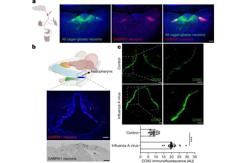
Through a series of experiments in mice, first author Na-Ryum Bin, HMS research fellow in the Liberles lab, identified a small population of neurons embedded in the glossopharyngeal nerve, which runs from the throat to the brain.
Importantly, he found that these neurons are necessary to signal to the brain that a flu infection is present and have receptors for lipids called prostaglandins. These lipids are made by both mice and humans during an infection, and they are targeted by drugs such as ibuprofen and aspirin.
Cutting the glossopharyngeal nerve, eliminating the neurons, blocking the prostaglandin receptors in those neurons, or treating the mice with ibuprofen similarly reduced influenza symptoms and increased survival.
Together, the findings suggest that these airway neurons detect the prostaglandins made during a flu infection and become a communication conduit from the upper part of the throat to the brain.
“We think that these neurons relay the information that there’s a pathogen there and initiate neural circuits that control the sickness response,” Liberles said.
The results provide an explanation for how drugs like ibuprofen and aspirin work to reduce flu symptoms—and suggest that these drugs may even boost survival.
The researchers discovered evidence of another potential sickness pathway, this one traveling from the lungs to the brain. They found that it appears to become active in the second phase of infection as the virus infiltrates deeper into the respiratory system.
This additional pathway doesn’t involve prostaglandins, the team was surprised to find. Mice in the second phase of infection didn’t respond to ibuprofen.
The findings suggest an opportunity for improving flu treatment if scientists are able to develop drugs that target the additional pathway, the authors said.
A foundation for future research
The study raises a number of questions that Liberles and colleagues are eager to investigate.
One is how well the findings will translate to humans. Although mice and humans share a lot of basic sensory biology, including having a glossopharyngeal nerve, Liberles emphasized that researchers need to conduct further genetic and other experiments to confirm that humans have the same neuron populations and pathways seen in the mouse study.
If the findings can be replicated in humans, it raises the possibility of developing treatments that address both the prostaglandin- and nonprostaglandin pathways of flu infection.
“If you can find a way to inhibit both pathways and use them in synergy, that would be incredibly exciting and potentially transformative,” Liberles said.
Bin is already delving into the details of the nonprostaglandin pathway, including the neurons involved, with the goal of figuring out how to block it. He also wants to identify the airway cells that produce prostaglandins in the initial pathway and study them in more depth.
Liberles is excited to explore the full diversity of sickness pathways in the body to learn whether they specialize for different types and sites of infection. Deeper understanding of these pathways, he said, can help scientists learn how to manipulate them to better treat a range of illnesses.
More information:
Stephen Liberles, An airway-to-brain sensory pathway mediates influenza-induced sickness, Nature (2023). DOI: 10.1038/s41586-023-05796-0. www.nature.com/articles/s41586-023-05796-0
Journal information:
Nature
Source: Read Full Article


