Home » Health News »
Researchers identify regions of the viral envelope of simian foamy viruses targeted by neutralizing antibodies
A recent study published in PLOS Pathogens characterized epitopes recognized by neutralizing antibodies (nAbs) from individuals infected with simian foamy virus (SFV).

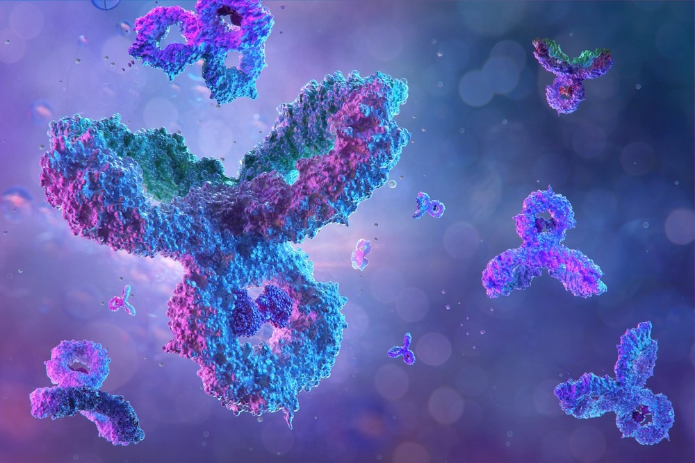
Study: Neutralization of zoonotic retroviruses by human antibodies: Genotype-specific epitopes within the receptor-binding domain from simian foamy virus. Image Credit: Corona Borealis Studio/Shutterstock.com
Background
SFVs are widespread in non-human primates (NHPs) and transmitted to humans through bites from infected NHPs. Most SFV-infected humans live in Central Africa. SFVs establish a persistent infection in humans with subclinical pathophysiological changes. Human-to-human transmission and severe illness have not been recorded to date, suggesting efficient control of replication and transmission in humans.
SFV infection induces envelope (Env)-specific nAbs that inhibit viral entry into susceptible cells in vitro. The Env has three subunits – leader peptide (LP) and surface (SU) and transmembrane (TM) subunits. Two env gene variants define the genotype of SFV strains. The variants differ at the SUvar region at the center of SU, which overlaps with the receptor-binding domain (RBD) and is targeted by nAbs.
The study and findings
In the present study, researchers determined the SFV SUvar epitopes recognized by nAbs from infected hunters in Central Africa. Neutralization assays were performed to define nAb epitopes in the presence of a recombinant SU competing with SU on the Env of foamy viral vector (FVV) particles.
Gorilla genotype I (GI) Env-derived constructs were poorly expressed, whereas GII Env-derived constructs were highly expressed. The immunoglobulin fragment crystallizable (Fc) domain was appended to the C-terminal end of SU to produce immunoadhesins for increased stability and yield. GI SU immunoadhesin expression was insufficient, whereas GII and chimpanzee genotype I (CI) SU immunoadhesins were readily expressed.
Therefore, epitopes recognized by GII- and GI-specific nAbs were tested using GII (GIISU) and CI (CISU) SU immunoadhesins, respectively, given the cross-neutralization of chimpanzee and gorilla SFV strains of the same genotype. Twelve plasma samples from SFV-infected hunters were used, including one from an individual infected with strains of two genotypes. Samples were tested by incubating with SU and adding to FVVs expressing matched Env.
CISU and GIISU inhibited the nAbs in genotype-matched samples but not in genotype-mismatched samples. The researchers noted that immunoadhesins did not completely block nAbs and speculated that this might be due to the lack of epitopes in the quaternary structure and the monomeric states. Therefore, they evaluated the effect of mammalian-type glycosylation and oligomerization of SUs in blocking nAbs.
GIISU constructs were produced in the presence of kifunensine (a mannosidase inhibitor) to prevent the addition of complex glycans. Some were additionally treated with endoglycosidase-H to remove all glycans from SU. The lack of complex glycans caused no change, but eliminating all glycans diminished the nAb blockade. Of the 11 glycosylation sites in SU, the N10 site knockdown mutant was as efficient as GIISU in inhibiting nAbs.
Further, other glycosylation sites were individually knocked down, except the N8 site critical for SU expression. These experiments indicated that most glycans, except those at the N7 site, were not involved in epitopes. Next, the researchers explored the location of nAb epitopes on SU subdomains. Deleting the central region of the RBD (RBDj) impaired nAb inhibition in samples from three subjects but was as efficient as GIISU against nAbs in one.
Furthermore, three loops (L2, L3, and L4) from the upper subdomain in GIISU were individually removed and evaluated. Deleting the L3 loop blocked all GII-specific samples except one, albeit with lower affinity. L2/L3 deletion had different plasma sample-dependent effects. In the lower subdomain, mutating residues in the heparan sulfate glycosaminoglycan-binding site (HBS) had a modest impact on nAb activity.
Further investigations identified the 345-353 loop and the α8-helix as major epitopes on GII RBD. The team applied a similar approach using GI-specific plasma and CISU constructs with mutations impacting GII-specific epitopes. They observed that GI-specific nAbs targeted glycans removed by endoglycosidase-H but not the complex glycans.
Deletion of RBDj or L3 in CISU failed to block nAbs. L2/L4 deletion had plasma-dependent effects, similar to GII counterparts. These data indicated that GI- and GII-specific nAbs target different epitopes. Moreover, most anti-SFV antibodies failed to recognize synthetic linear peptides spanning the upper RBD subdomain loops.
All SU constructs were evaluated for binding to human HT1080 cells highly susceptible to gorilla/chimpanzee SFVs. Glycan removal in SUs enhanced cell binding, whereas RBDj deletion decreased binding capacity, which was more potent in GII than CI SU. The mutant SUs that showed cell binding were tested for nAb inhibition. Overall, only one mutant SU failed to bind to cells and inhibit nAbs.
Conclusions
The researchers uncovered epitopic regions across SFV SUvar recognized by GII-specific nAbs from infected humans, with at least one recognized by GI-specific nAbs. They hypothesized that anti-SFV nAbs mainly target sites critical for Env trimerization, conformational changes leading to membrane fusion, and those involved in cell binding.
- Dynesen, L. et al. (2023) "Neutralization of zoonotic retroviruses by human antibodies: Genotype-specific epitopes within the receptor-binding domain from simian foamy virus", PLOS Pathogens, 19(4), p. e1011339. doi: 10.1371/journal.ppat.1011339. https://journals.plos.org/plospathogens/article?id=10.1371/journal.ppat.1011339
Posted in: Medical Science News | Medical Research News | Disease/Infection News
Tags: Antibodies, Cell, Chimpanzee, Gene, Glycan, Glycans, Glycosaminoglycan, Glycosylation, Helix, Immunoglobulin, Membrane, Peptides, Receptor, Viral Vector, Virus

Written by
Tarun Sai Lomte
Tarun is a writer based in Hyderabad, India. He has a Master’s degree in Biotechnology from the University of Hyderabad and is enthusiastic about scientific research. He enjoys reading research papers and literature reviews and is passionate about writing.
Source: Read Full Article


