Home » Health News »
Study reveals journey of immune cells in developing zebrafish

Microglia, the immune cells of the brain, form the first line of defense against neurodegenerative diseases and traumatic brain injuries. They maintain brain homeostasis, the stable condition necessary for survival, by acting like tiny vacuum cleaners—congregating at damaged areas, removing dead, infected, or injured brain cells and tidying up unnecessary synapses. However, microglia don’t originate in the brain, rather their precursor form travels there during development from their place of origin, another section of the embryo called the peripheral mesoderm. Many questions around this process, such as how the microglial precursors find their way, are currently unanswered. And, given how important these cells are for brain homeostasis in all animals, including humans, answering these questions could have a myriad of health benefits.
“Microglia have been implicated in several neurobiological diseases such as Alzheimer’s. Revealing how they work could shed light on these diseases,” stated Prof. Ichiro Masai who leads the Developmental Neurobiology Unit at the Okinawa Institute of Science and Technology Graduate University (OIST). “Microglia are found throughout the brain, but our most recent paper looks at how they colonize the retina—the neural tissue of the eye, which is one of the first brain regions to accommodate these cells.”
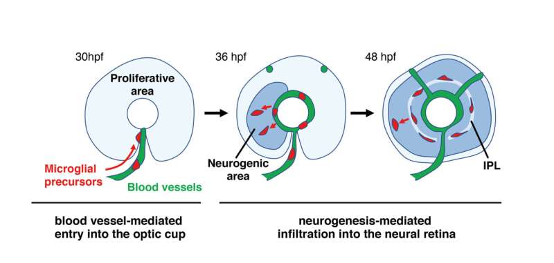
Reporting in eLife, Prof. Masai and former OIST Ph.D. student Dr. Nishtha Ranawat revealed that, to successfully colonize the retina, microglia require two processes to have occurred—the blood vessels must have formed inside the eye, and neurogenesis, the process of neuron formation, must have begun within the retina.
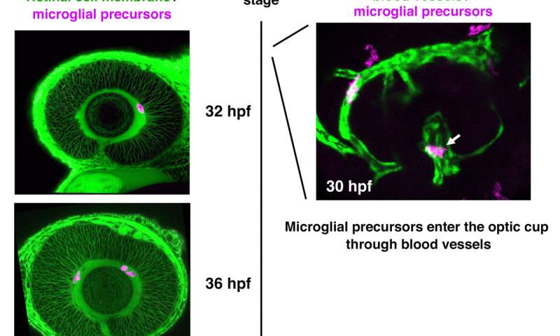
To uncover these requirements, the researchers imaged transparent embryonic zebrafish from 24 hours after fertilization to 60 hours after fertilization. They tagged the microglia precursors with fluorescence, which allowed them to be tracked. From their original place, these precursors first traveled to the yolk, and started migrating towards the various brain regions from there.

“The developing brain is very packed tissue,” explained Prof. Masai. “It’s hard to imagine how the microglia enter it and move around inside. But we found that they use the blood vessels as pathways to enter the eye.”
However, the journey doesn’t end there. Once the microglia arrive at the entrance of the retina, they remain associated with the blood vessels between the retina and the lens until another process begins—neurogenesis. Neurogenesis is the process of neuron formation. The researchers found that microglia could only infiltrate areas of the retina where neurogenesis was occurring. At around 60 hours after fertilization, the microglia were in place, spread throughout the retina.
To further determine that the blood vessels and neurogenesis were important components for microglia migration, the researchers hindered, in one experiment, the blood vessels from forming and, in another experiment, neurogenesis from occurring. In both cases, microglia were unable to enter the retina.
Source: Read Full Article


