Home » Health News »
Study demonstrates minimally invasive, aerosolized vaccine candidate to combat SARS-CoV-2
Macrophages play a crucial part in inducing both innate and adaptive immunity in the human body. Their major roles include capacity for antigen presentation to T-helper cells, which induce a cascade of responses, recruit inflammatory cytokines and mediate adaptive immune responses.
 Study: Inhalable SARS-CoV-2 Mimetic Particles Induce Pleiotropic Antigen Presentation. Image Credit: Josep Suria/Shutterstock
Alveolar macrophages are one of the two major classes of macrophages in the human lungs which are in close contact with the alveolar epithelium. They serve as the first line of defence against invading pathogens entering the body through noxious stimuli. These immune cells alter their polarization on stimulation, from the naïve state (Mø) to pro-inflammatory (M1) or anti-inflammatory (M2) phenotypes, combatting invading pathogens. These characteristics make them ideal targets for vaccination.
Background
Angiotensin-converting enzyme 2 (ACE2), the key binding receptor of the viral spike protein in severe acute respiratory syndrome coronavirus (SARS-CoV-2), is expressed abundantly on the activated macrophageal surfaces. This marks the first step of coronavirus (COVID-19) pathogenesis.
To this end, activated alveolar macrophages generate contradictory biologic effects, initiating both important antiviral defence mechanisms on one hand, and the “Trojan horse” macrophage hosts on the other, that promotes binding of SARS-CoV-2 within the pulmonary parenchyma. Additionally, virus-infected macrophages may circulate and spread the infection to distal organs and cause systemic infection. This phenomenon is referred to as the “macrophage paradox”.
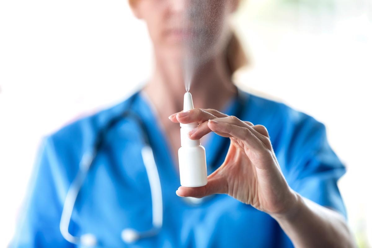
Considering the importance of alveolar macrophages in Covid-19 pathogenesis, researchers speculated the targeted delivery of SARS-CoV-2 antigens to these cells via inhalable vaccines in a recent study published in Biomacromolecules. They hypothesized that such a non-invasive immunization process may yield potent prophylactic mucosal immunity among the masses.
Study details
Researchers developed inhalable coronavirus mimetic particles (CoMiP) designed to be rapidly internalized and identified by macrophages to preferentially deliver viral antigens and equip the immune cells to make antibodies. They encoded plasmids within carriers as an initial model system. They mimicked the basic structure of the SARS-CoV-2 virion and packaged the nucleic acid-equivalent structures of CoMiP within a peptide envelope coated with a glycosaminoglycan and hyaluronic acid. This versatile platform ensured pleiotropic transfection of macrophages and lung epithelial cells with multiple antigen-encoding plasmids.
Researchers developed a model GFP-encoding plasmid to optimize the formulation of CoMiP in the laboratory. The GFP plasmid was first mixed with PLL to form nanoscale polyplexes. SEM and zeta potential measurements confirmed the formation of 100nm-radius, amorphous, cationic complexes. Adding hyaluronic acid made for spiky nanoparticles with an approximate size of∼150 nm, with similar surface topography as the SARS-CoV2 virion.
Researchers used human THP-1 monocytes polarized to an Mø macrophage phenotype,mimicking naïve alveolar immune cells to evaluate cellular phagocytosis of the eGFP constructs (eGFP-encoding CoMiP (CoMiPGFP)). Flow cytometry-based analysis showed that that >20% of treated THP-1 macrophages were successfully transfected.
Researchers then selected a panel of plasmids encoding for all four key SARS-CoV-2 structural proteins, including spike (S), envelope (E), membrane (M), and nucleocapsid (N). Two transgene candidates were chosen to either encode the receptor-binding domain (SRBD36) or the full-length (S) spike protein. Quantification assays demonstrated successful encapsulation of 17−47% of available plasmid cargo into the CoMiPGFP constructs.
Researchers then optimized the formulation of S-plasmid loaded CoMiP through several in vitro assays using tolerance studies performed in CoMiP-treated endothelial (HUVEC) and macrophage (THP1) cell lines. Follow-up studies evaluated the transfection of RAW264.7 murine macrophages 72 h after treatment with each CoMiP formulation.
As a next step, researchers explored the efficacy of the inhalable CoMiPS vaccines, by injecting C57BL/6 mice via the intranasal instillation of a 40 μL volume of 29 μg/mg CoMiPS in sterile saline. Healthy control animals received a similar volume of saline water as a placebo.
All animals were boosted with the same concentration 2 weeks later. Systemic immune responses were then evaluated via ELISA performed on serum samples collected from animals 14 and 28 days, each, after initial vaccination and subsequent booster, respectively.
Quantification assays of S-protein-specific serum immunoglobulin G (IgG) showed no statistically significant change in systemic antibody levels between CoMiPS-treated animals and controls at both sampling time points. The main key was assessing the mucosal immune responses (IgA levels), for which bronchoalveolar lavage fluid (BALF) was collected from immunized mice 30 days after vaccination.
While there was a significant increase in the total IgA for mice vaccinated with CoMiPS, no statistically significant difference in S-protein specific IgA was detected between the vaccinated and control animals. Researchers then conducted histology-based analysis on lung tissues collected at the end of the vaccination period.
The samples indicated no overt signs of pulmonary inflammation, which would otherwise show extensive cell infiltration, septal thickening, and collapsed airways in infected lung tissues. This observation was particularly important as the low-molecular-weight hyaluronic acid (100 kDa) used to construct the CoMiP carrier could potentially have stimulated leukocyte infiltration to cause counter airway inflammation, hyperresponsiveness, and tissue remodelling.
Implications
This study showed CoMiP carriers as potential, versatile, aerosolizable vaccine candidates if worked on with continued optimization and refinement. This would be a big leap towards minimally invasive immunization therapies for SARS-CoV-2 and potentially other respiratory viruses that would elicit local immunity at the primary site of infection.
- Lawanprasert A, Simonson AW, Sumner SE, Nicol MJ, Pimcharoen S, Kirimanjeswara GS, et al. (2022). Inhalable SARS-CoV-2 Mimetic Particles Induce Pleiotropic Antigen Presentation. Biomacromolecules. doi: https://doi.org/10.1021/acs.biomac.1c01447 https://pubs.acs.org/doi/10.1021/acs.biomac.1c01447
Posted in: Medical Science News | Medical Research News | Disease/Infection News
Tags: ACE2, Airway Inflammation, Angiotensin, Angiotensin-Converting Enzyme 2, Antibodies, Antibody, Antigen, Anti-Inflammatory, Cell, Coronavirus, Coronavirus Disease COVID-19, covid-19, Cytokines, Cytometry, Efficacy, Enzyme, Flow Cytometry, Glycosaminoglycan, Histology, Horse, Immune Response, immunity, Immunization, Immunoglobulin, in vitro, Inflammation, Laboratory, Leukocyte, Lungs, Macrophage, Membrane, Nanoparticles, Nucleic Acid, Phagocytosis, Phenotype, Placebo, Plasmid, Protein, Receptor, Respiratory, SARS, SARS-CoV-2, Severe Acute Respiratory, Severe Acute Respiratory Syndrome, Spike Protein, Syndrome, Transfection, Transgene, Vaccine, Virus

Written by
Sreetama Dutt
Sreetama Dutt has completed her B.Tech. in Biotechnology from SRM University in Chennai, India and holds an M.Sc. in Medical Microbiology from the University of Manchester, UK. Initially decided upon building her career in laboratory-based research, medical writing and communications happened to catch her when she least expected it. Of course, nothing is a coincidence.
Source: Read Full Article


