Home » Health News »
Novel technique decodes mechanisms controlling executive functions of the primate brain

The human brain is a wonderfully enigmatic organ, helping to juggle multiple tasks efficiently to help us get through a long day. This feature, called executive function, seats primates like us at the pinnacle of evolution. The prospect of losing the spectacular flow of neural information in our brains because of an accident or disease is, thus, unnerving. In the event of such an unfortunate occurrence, to restore the brain to its previous working condition with full functionality—to reboot it, so to speak—would need a better understanding of the specific neural pathways that rely on working memory and decision-making—two important executive functions.
To achieve this objective, a group of researchers from National Institutes for Quantum and Radiological Science and Technology (QST), Japan, developed a technique they call “imaging-guided chemogenetic synaptic silencing” to decipher the specific neural pathways involved in high-order executive functions. In a pioneering study published in Science Advances, they now report successfully delineating specific neural pathways involved in working memory and decision-making using this technique.
The group, led by Dr. Takafumi Minamimoto from the Department of Functional Brain Imaging, QST, focused on studying the dorsolateral part of the prefrontal cortex (dlPFC) in the monkey brain to apply their technique, and further identify the neural pathways of interest. They chose this brain region as it is partially responsible for controlling executive functions and is only present in primates.
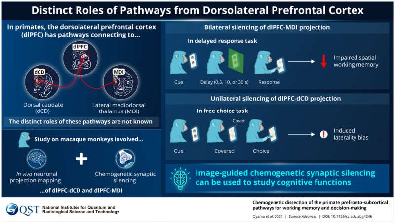
Importantly, the role of dlPFC is supported by brain regions like the dorsal caudate (dCD) and lateral mediodorsal thalamus (MDl). Regarding this intricate association, Dr. Kei Oyama, first author of the study, says, “The primate prefrontal cortex (PFC), especially its dorsolateral part, is well known to serve as the center of higher-order executive functions; it is uniquely developed in primates and underlies their distinctive cognitive abilities. These functions, however, do not solely rely on dlPFC neurons but also on their cooperative interactions with subcortical structures, including the dorsal caudate (dCD) nucleus and lateral mediodorsal thalamus (MDl).”
Next, the researchers wanted to identify the mechanisms for working memory and decision-making. Given that the dlPFC, MDI, and dCD neurons are connected, they selectively silenced specific neuronal synapses to disrupt the flow of information to achieve dlPFC-dCD and dlPFC-MDl projections, either unilaterally (involving just one side of the brain), or bilaterally (involving both sides). They made the dlPFC neurons express designer receptors exclusively activated by designer drugs (DREADDs). Further, the monkeys involved in the study were analyzed for behavioral changes to understand the effect of chemogenetic silencing.
Interestingly, the researchers observed that silencing the bilateral dlPFC-MDl projections in the monkeys, but not their dlPFC-dCD projections, caused problems in working memory related to their surroundings. On the contrary, silencing their unilateral dlPFC-dCD projections, but not their unilateral dlPFC-MDl projections, altered their preference in decision-making. These results reveal that the two higher-brain functions, working memory and decision-making, are controlled by different neural pathways linking specific brain areas.
Source: Read Full Article


